

 +852 2866 8995
 General Enquiry: cs@medikonia.com | Support: support@medikonia.com


All products from TargetMol are for Research Use Only. Not for Human or Veterinary or Therapeutic Use.
Biological Description
Description: 3-Methyladenine (3-MA) is a selective PI3KV inhibitor, and the IC50s against ps34 and PI3Kγ were 25/60 μM in HeLa cells, respectively.
Targets&IC50: PI3Kγ:60 μM (in HeLa cells), Vps34:25 μM (in HeLa cells)
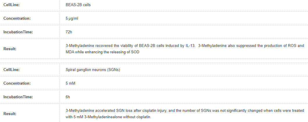
In vitro: Although 3-MA shows some limited Vps34 preference in vitro, with an IC50 of 25 μM for Vps34 as compared with 60 μM for PtdIns3Kγ it is typically employed in cellular studies at a concentration of 10 mM, which can inhibit all PtdIns3Ks. The treatment of cerebellar granule cells with either 20 or 10 mM 3-MA prevented both autophagosome proliferation and cell death, without affecting neuronal morphology nor protein synthesis. Treatment with 5 mM 3-MA decreased the percentage of glucose-starved HeLa cells displaying GFP-LC3 puncta to 23%. Treatment of HeLa cells with 2.5 mM or 5 mM 3-MA for one day did not affect cell viability, whereas treatment with 10 mM 3-MA for one day caused a 25.0% decrease in cell viability. Treatment of cells with 2.5, 5 or 10 mM 3-MA for two days caused 11.5%, 38.0% or 79.4% decrease in viability, respectively.
In vivo: In severe acute pancreatitis (SAP) group, the pathological change increased with time after modeling. Pathological change of the pancreas tissue in 3-methyladenine group was milder than those in the SAP group at both 12 and 24 h. 3-MA pretreatment significantly aggravated neurological symptoms when compared with the SAH + vehicle group. a large number of dying neurons from the SAH + 3-MA group showed cell shrinkage, chromatin condensation at the nuclear membrane and nuclear and cellular fragmentation, which suggest that the neurons were undergoing apoptotic cell death.
Cell Research: Cells were seeded in an 8-well coverglass-bottomed chamber for 24 hours (6×10^3 cells per well). Images were acquired automatically at multiple locations on the coverglass using a Nikon TE2000E inverted microscope fitted with a 20× Nikon Plan Apo objective, a linearly-encoded stage, and a Hamamatsu Orca-ER CCD camera. A mercury-arc lamp with two neutral density filters (for a total 128-fold reduction in intensity) was used for fluorescence illumination. The microscope was controlled using NIS-Elements Advanced Research software and housed in a custom-designed 37°C chamber with a secondary internal chamber that delivered humidified 5% CO2. Fluorescence and differential interference contrast images were obtained every 10 min for a period of 48 hours. To analyze live cell imaging movies, the time-lapse records of live cell imaging experiments were exported as an image series and analyzed manually using NIS-Elements Advanced Research software. The criteria for analyses were described previously, and lagging chromosomes in prometaphase were defined as the red fluorescence-positive materials that lingered outside the roughly formed metaphase plate for more than 3 frames (30 min).
Animal Research: All rats were fasted for 12 h with free access to water prior to operation. After anesthesia by intraperitoneal (i.p.) injection of 2% sodium pentobarbital (0.25 mL/100 g), they were laid and fixed on the table, routinely shaven, disinfected, and draped. The rat SAP model was induced by 0.1 mL/min speed uniformly retrograde infusion of a freshly prepared 3.5% sodium taurocholate solution (0.1 mL/100 g) into the biliopancreatic duct after laparotomy. Equivalent volume of normal saline solution was substituted for 3.5% sodium taurocholate solution in the sham-operation (SO) control group. The incision was closed with a continuous 3-0-silk suture, and 2 mL/100 g of saline was injected into the back subcutaneously to compensate for the fluid loss. 180 rats were randomly divided into four groups: (1) Acanthopanax treatment group (Aca group, n = 45) where the rats were injected with 0.2% Acanthopanax injection at a dose of 3.5 mg/100 g 3 h after successful modeling via the vena caudalis once, knowing that this dosage was effective as proven in our previous experiment; (2) 3-Methyladenine treatment group (3-methyladenine group, n = 45) where the rats were injected with 100 nmol/μL 3-methyladenine solution at a dose of 1.5 mg/100 g 3 h after successful modeling via the intraperitoneal route once, knowing that this dosage was effective as proven in the literature; (3) SAP model group (SAP group, n = 45) where these rats received an equivalent volume of the normal saline instead of Acanthopanax injection 3 h after successful modeling via the vena caudalis once; (4) SO group (control, n = 45) where these rats received an equivalent volume of the normal saline instead of Acanthopanax injection 3 h after successful sham-operation via the vena caudalis once. The 45 animals in each of the four groups were equally randomized into 3, 12, and 24 h subgroups for postoperative observations.
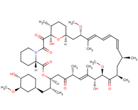
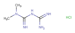
Chemical Properties
Molecular Weight: 149.157
Formula: C6H7N5
CAS No.: 5142-23-4
Storage & Solubility Information
Storage
Powder: -20°C for 3 years
In solvent: -80°C for 2 years
Solubility Information
DMSO: 3 mg/mL (20.11 mM), warmed
Ethanol: 4 mg/mL (26.81 mM)
H2O: 8 mg/mL
( < 1 mg/ml refers to the product slightly soluble or insoluble )
Catalog #: T1879
Please email us at cs@medikonia.com for any enquiry. To place an order, please include the catalog number(s) of the product(s) in the email.
References and Literature
General Enquiry: cs@medikonia.com | Support: support@medikonia.com
+852 2866 8995
Unit 1005-1006, 10/F, Tower I, Grand Century Place, 193 Prince Edward Road West, MongKok, Kowloon, Hong Kong


Powered by Medikonia Limited 2023