

 +852 2866 8995
 General Enquiry: cs@medikonia.com | Support: support@medikonia.com


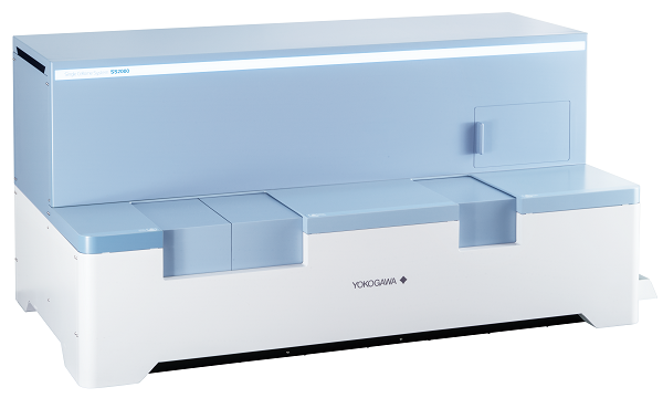
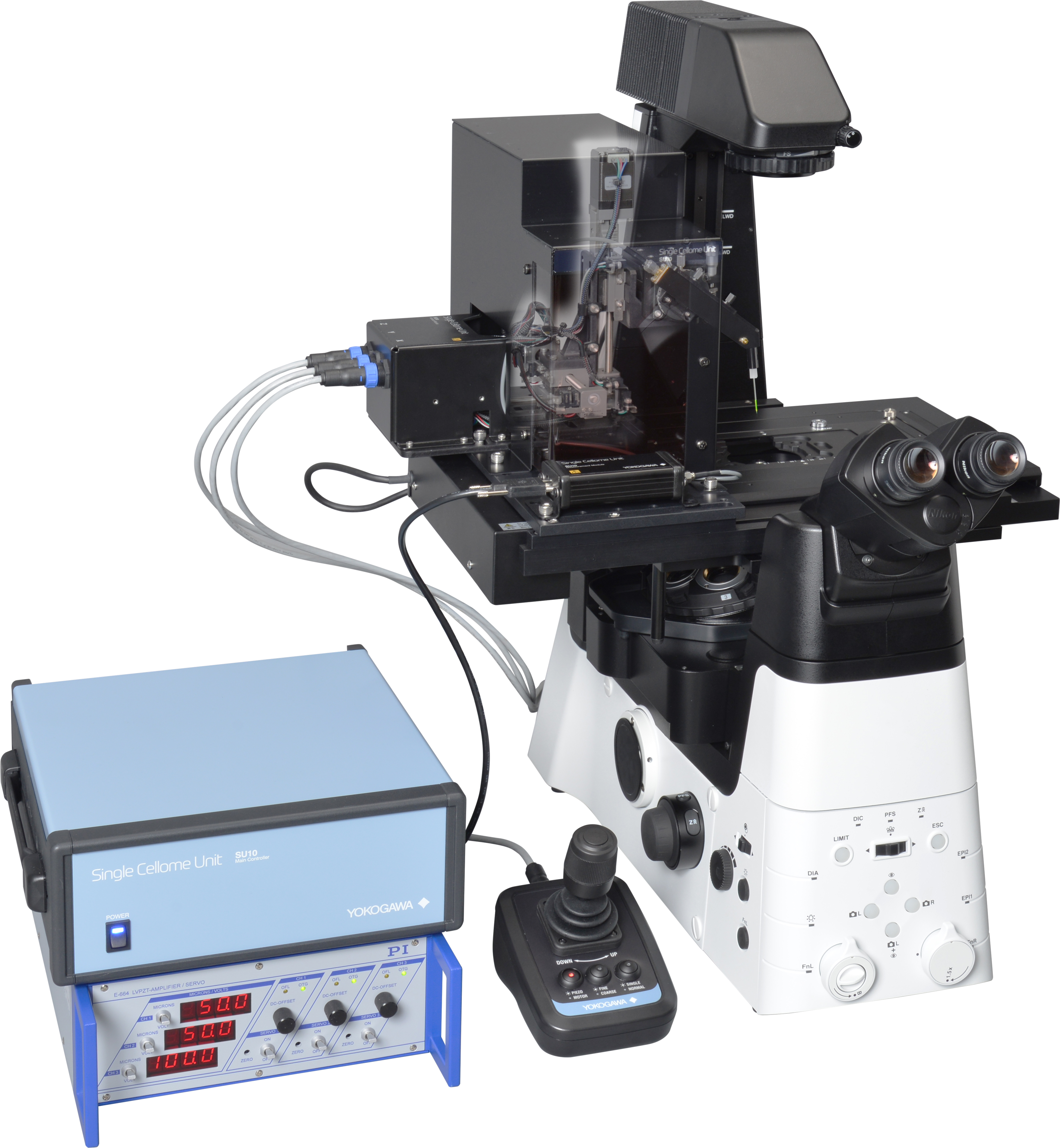
Yokogawa Single Cellome™ System SS2000
The SS2000 is a system that automatically samples specific regions of cells or whole cells at the single-cell level while imaging cells in culture with a confocal microscope. Because cells in culture do not need to be detached, positional and morphological information is maintained. #Subcellular Sampling System


 Unlike existing cell isolation devices, the SS2000 can not only isolate a whole-cell but also sample only the target site inside the cell. It is possible to sample cytoplasm and regions containing target organelles selectively.
After staining of HeLa cell nuclei (blue), cytoplasm (green), and mitochondria (red), a mitochondria-rich region (arrow) of the cytoplasm was sampled.
 Since only the target cells can be sampled without detaching the cells in culture, it is possible to sample while maintaining positional and morphological information.
Normal MDCK cells and green fluorescent-labeled abnormal MDCK cells were co-cultured at a 50:1 ratio. A normal cell adjacent to the abnormal cell exhibiting fluorescent signals (arrows) was sampled.
Samples can be collected on PCR plates and microplates and collect multiple samples in the same well. Samples can also be taken while being held in the glass tip without being ejected. The collection site has a cooling function to suppress sample degradation and an incubator function to maintain the culture environment. These samples can be used for genetic analysis, mass spectrometry, and single-cell cloning.

The SS2000 utilizes live-cell imaging products developed by Yokogawa. High-speed, high-resolution 3D imaging is possible using our unique confocal microscope technology. Samples can be taken from targeted cells under a confocal microscope in an incubator environment. Time-lapse photography is also possible, allowing dynamic changes in the target cell to be captured. Since it is possible to record moving images during sampling and images before and after sampling, it is possible to compare the results of analysis of collected samples with cell imaging data.
Target cells and sampling positions can be automatically selected by image analysis. (Targets can be automatically selected as shape of cells, size of nuclei, density of organelles etc.)


| Automatic sampling functions | Tip diameter | 3μm, 5μm, 8μm, 10μm |
|---|---|---|
| Incubator loader environment | 37℃, 5%CO2, humidified | |
| Collection loader environment | 37℃, 5%CO2, humidified (for culture) / 4℃ (for cooling) | |
| Collection loader compatible vessels | 96-well PCR plate (0.1mL, 0.2mL) Multiwell culture plate (96well) | |
| Postioning precision of sampling | XYZ axial designated resolution: 0.1μm | |
| Imaging functions | Confocal scanning method | Microlens enhanced dual wide Nipkow disk confocal |
| Incubator loader compatible vessels | When sampling cell: φ35mm dishes *1 Microplate (6well, 24well, 96well) |
|
| When observing cell: φ35mm dishes *1 Microplate (6well, 12well, 24well, 48well, 96well, 384well, 1536well) Slideglass *2 |
||
| Excitation laser wavelength | 405, 488, 561, 640nm (Uniformizer installed) | |
| Emission filter | Filter size: φ25nm, Maximum slot number: 10 (Electric switching), Adjacent switching speed: 100msec | |
| Transmission illumination | Bright-field, LED source | |
| Objective lens | Dry lens:4x, 10x, 20x, 40x Long-working distance lens:20x, 40x Note that only the 40x dry lens can be used for cell sampling. |
|
| Z focus | Electric Z motor, designated resolution:0.1μm | |
| Electric stage | XYZ axial designated resolution:0.1μm | |
| Autofocus | Laser autofocus | |
| Camera | sCMOS camera 2,000 x 2,000pixel Pixel size:6.5 x 6.5μm | |
| Other | Special purpose workstation | Workstation for sampling, measurement, analysis, 24 inch display x2 |
| Measurement software | Measurement functions (2D, 3D, Time-lapse, Map imaging), Viewing measurement and sampling data, Reporting functions (Image data, Video data), Whole cell sampling, Intracellular component sampling | |
| Analysis software | Analysis functions (3D, Tile, Label-free, Texture analysis, Deep Learning, Gating), 3D viewer, Graphing functions, Reporting functions(Image data, Video data, EC50, IC50, Z'-factor) | |
| External dimensions, Weight | Main unit: W1,217 x D643 x H595 mm, 145kg Utility box: W275 x D432 x H298 mm, 18kg Gas mixer: W275 x D432 x H298 mm, 10kg Special purpose workstation: W172 x D471 x H414 mm, 14kg Display: W531 x D500 x H166 mm, 5.6kg |
|
| Operating environment | Temperature: 15 to 30℃ Humidity: 30 to 70%RH no condensation |
|
| Power consumption | Main unit, Utility box and Gas mixer: 1,200VAmax Workstation: 950VAmax Display: 42VAmax x 2 |
|
| Data formats (Measurement software) | Captured images : 16bit TIFF (OME-TIFF, TIFF) Output image data : TIFF, PNG, JPEG Output video data : WMV, MPEG4 |
|
|
Data formats (Analysis software) |
Numeric data: CSV Output image data: TIFF, PNG, JPEG Output video data: WMV, MPEG4 |
*1 A sample holder is required, and with it, up to 3 samples can be installed.
*2 A sample holder is required, and with it, up to 4 samples can be installed.
For more information or ordering the products, please email us at cs@medikonia.com.
General Enquiry: cs@medikonia.com | Support: support@medikonia.com
+852 2866 8995
Unit 1005-1006, 10/F, Tower I, Grand Century Place, 193 Prince Edward Road West, Mong Kok, Kowloon, Hong Kong


Powered by Medikonia Limited 2023