

 +852 2866 8995
 General Enquiry: cs@medikonia.com | Support: support@medikonia.com


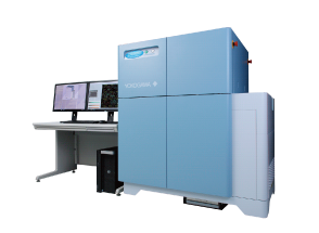
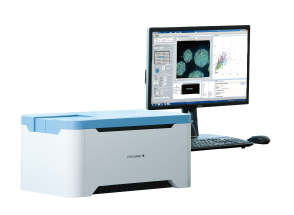
Yokogawa CellVoyager CV8000 High-Content Screening System
Real time confocal, label-free imaging
Within the drug development market, demands on high content analysis systems for drug efficacy evaluation are increasing in accordance with the needs for cell-based assay and phenotypic screening. In order to increase screening efficiency, devices with higher speeds (higher throughput) are required.
On the other hand, in order to bridge the “valley of death” of the drug development process, the quality of screening hits must be increased.
This requires the construction of more complex evaluation systems that utilize multifaceted parameters via 3D cultivation systems, live-cell imaging and higher detail image analysis.
In current drug development research, determining how to implement throughput screening and complex evaluation system screening in parallel is an important issue.
| Excitation laser wavelength | 405nm、445nm、488nm、561nm、640nm |
| Illumination source | Laser |
| Objective lens | 2x to 60x (Dry, Phase contrast, Water immersion, Long working distance) |
| Camera | High-sensitivity sCMOS camera (up to 4 units) |
| Autofocus | Laser-based mode, image-based mode |
| Software | CellPathfinder、CellLibrarian |
The CellVoyager CV8000 is a high-end, high content analysis system that solves this contradictory screening challenge.
Through the combination of a proprietary Yokogawa high speed confocal scanner, water immersion lens, up to four high field-of-vision cameras, a microscopic stage with cell cultivation environment, and an integrated robotic pipetter, we have realized not only high throughput, high-resolution imaging, but also phenotypic screening via a more complex evaluation system.
In addition, our specialized analysis software, CellPathfinder, uses deep leaning and machine learning to recognize target objects with high accuracy, supporting you from image analysis to results display using graphs.
Real time confocal, label-free imaging

Stage incubator included as standard. Realization of non-stop, long-duration observation (3 days +) via humidity, temperature and CO2 control.
Left:Before incubation Right:After 68 hours incubation

Drug addition during imaging is made possible by an integrated robotic pipetter with disposable tips.
Ideal for kinetic experiments involving the observation of high speed phenomena.
Left:Before incubation Right:After stimulation

Yokogawa’ s spinning disk confocal technology excels in imaging of samples with depth, such as 3D culture samples where clear and quick imaging is difficult, allowing for evaluation close to in-vivo quality.
Left:Original image Right:Recognition image

Recognition and analysis can be performed by taking bright field images from several Z positions and creating a CE bright field image using the included CellPathfinder analysis software. Analysis accuracy is further enhanced via the new Deep Learning option.
Left:CE Bright Field Right:Cell Recognition image
Observe cells as they are -Dual spinning disk confocal system-
 A Yokogawa proprietary multi-scan method utilizing approximately 1,000 laser beams on the observation region and tandem disks rotating at high speed. The disks comprise a pinhole array disk with approximately 20,000 pinholes arranged in an equal pitch spiral pattern, and a microlens array disk that focuses the excitation light laser into individual pinholes. Not only does this allow high speed imaging, but it also largely prevents phototoxicity and fluorescence photobleaching.
 Deeper, clearer observation -Pinhole disk exchanger-
Two different types of pinhole disks (25/50μm) can be used, according to the sample. For thick samples, reducing the pinhole diameter allows for higher confocality, shaper images. For dark samples, increasing the pinhole diameter allows for brighter images.
Organoid imaging example Upper:25μm pinhole Lower:50μm pinhole

The optical system configuration can be selected according to the purpose. A single 96-well plate can be imaged in four colors in one minute by attaching four high- sensitivity wide -field sCMOS cameras. The system is also compatible with FRET and CellPainting assay.

Water immersion lenses excel in capturing high-resolution images of cells within a liquid. The CV8000 can be equipped wit h a 40x or 60x water immersion objective lens. Our 40x lens is a particularly unique lens capable of highly advanced spherical aberration correction, allowing for the capture of bright high-resolution images over a full wide -field. The lens water supply is also completely automated. This equipment makes high throughput screening via water submersion lens possible.
 Capturing live cell movement -High-precision incubator and robot pipetter-
The stage incubator features an airtight construction, managing humidity, temperature and CO2 levels. The integrated robotic pipetter conducts the following process fully automatically: tip pickup → reagent collection from the reagent plate → reagent addition to the sample plate → tip disposal. Not only can images be rapidly obtained before and after reagent instillation, but it’s also possible to add reagents to single wells multiple times, and adjust the addition speed etc., broadening the range of dynamics observation via reagent instillation.
HeLa cells were seeded in a 96 well plate at a density of 500 cells per well, and cultured for 24 hours. The well plate was then placed in the internal stage incubator and cell culturing was conducted for 72 hour s, and the total area (hereinafter Total Area) occupied by cells was analyzed. As a result, minimal unevenness in cell multiplication was observed across the 96-wells (excluding the four corner wells) when compared to a regular CO2 incubator.

Cell multiplication comparison with regular CO2 incubator after 72hr incubation(n=3)
The values represent the following: CV8000 Total Area after 72hrs / Total Area at 0hrs (hereinafter Total Area ratio) / CO2 incubator Total Area ratio x 100.
(Numbers near to 100 me an that cell multiplication was approximately equal for the CV8000 and CO2 incubator.)
Cell multiplication near to that of the CO2 incubator was verified, excluding the four corner wells.

Cell multiplication curves for each well of a 96-well plate
Cell multiplication was low in the four corner wells; however, it continued in the other wells.

Total Area ratio after cultivation start (24, 48 and 72 hours) (n=3)
Excluding the four corner wells, even after 72 hours, there were no large differences in cell multiplication.
The low variation in cell multiplication speed across the wells can 24 hours 48 hours 72 hours be seen.
Centralized process management, from the cultivation environment, to transfer, imaging, analysis and data management.
We offer optimum systems in response to our customers’ needs.

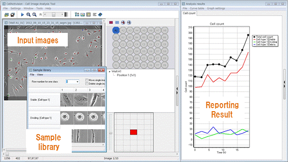
The software analyzes image data captured with the CV8000, creates graphs and exports various data. Beginner and expert users alike can take full advantage of the software, thanks to an abundance of templates and flexible protocol editing capability. CE bright field and machine-learning functionalities make label-free analysis possible. The new Deep Learning option has also been added, largely improving cell recognition accuracy.

Simply follow the flow displayed at the top of the screen. The analysis menu has easy-to-understand icons. Simply click the desired menu item and the protocol will load.

Computed numeric data can be displayed in a variety of ways. Graph plots and cell images are linked, making for easy result verification and inquiry.

Machine-learning also provides bias-free digitization of visually-evaluated experiments. Automatic recognition is made possible simply by clicking the shape you want the software to learn.

Eliminates the time, cost and influence on cells associated with cell labelling. Even higher precision classification is made possible through combination with deep learning.
For more information or ordering the CellVoyager CV8000 High-Content Screening System in Hong Kong or China, please email us at cs@medikonia.com
General Enquiry: cs@medikonia.com | Support: support@medikonia.com
+852 2866 8995
Unit 1005-1006, 10/F, Tower I, Grand Century Place, 193 Prince Edward Road West, Mong Kok, Kowloon, Hong Kong


Powered by Medikonia Limited 2023