

 +852 2866 8995
 General Enquiry: cs@medikonia.com | Support: support@medikonia.com


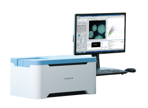
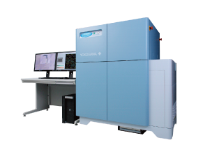
Yokogawa CellVoyager™ CQ1 Benchtop High-Content Analysis System
CellVoyager™ CQ1 enables 3D imaging and quantification of live cell clusters, such as spheroids within a 3D culture vessel, as they are, keeping the cells intact. CellVoyager™ CQ1 exports feature data in general formats which are readable by various third-party software for advanced data analysis. It is possible to construct a fully customized CellVoyager™ CQ1-based system by integrating with external systems*1, via robot for culture dish handling.
| Excitation laser wavelength | 405 nm, 488 nm, 561 nm, 640 nm |
| Illumination source | Laser |
| Objective lens | 2x to 60x (Dry, Phase contrast, Long working distance) |
| Camera | High-sensitivity sCMOS camera |
| Autofocus | Laser autofocus, Software autofocus |
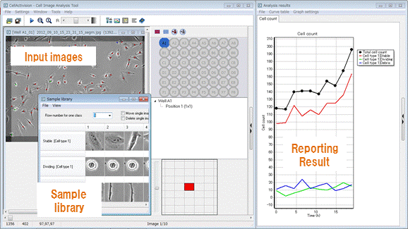
| Software | CellPathfinder |
 Enables Measurement of Spheroids, Colonies, and Tissue Sections
| CQ1 | General fluorescent imaging | Flow cytometry | |
|---|---|---|---|
| Cell removal/suspension treatment | Not necessary | Not necessary | Necessary |
| Cell image confirmation | Possible | Possible | Not possible |
| Display feature data and graphs in real-time with imaging | Possible | Depends on devices | Possible |
| 3D data measurement | Possible | Not possible | Not possible |
| Time-lapse | Possible | Not possible | Not possible |
*1 Option
*2 Contact to CQ1 partner for more information
Compact design contains fully integrated multiple functions to offer an easy-to-handle confocal imaging system, without a need for complicated system integration. You only need to set a sample and run the software. A user-friendly interface and versatile functions support your measurement and analysis.

A Nipkow spinning disk containing about 20,000 pinholes and a subsidiary spinning disk containing the same number of microlenses to focus excitation laser light into each corresponding pinhole are mechanically fixed on a motor, and very rapidly rotated. As a result, a high-speed raster scan of the excitation lights on the specimen can be achieved. The pinhole and microlenses are arranged on each disk in our proprietary design to optimize the raster scan. Multi-beam scanning not only increases scanning speed but also results in significantly lower photobleaching and phototoxicity because multi-beam excitation needs only a low level of laser power on the specimen to fully excite fluorescence.


The software learns the features of the sample objects collected by users.


Label-free Analysis

*1 Optional software
*2 Digital phase contrast

| Item | Specifications |
|---|---|
| Optics | Microlens enhanced dual wide Nipkow disk confocal, transmitted illumination |
| Laser/Filter | Laser: Choose 2-4 lasers from 405/488/561/640 nm, 10-position filter wheel (built-in) |
| Camera | sCMOS 2000 × 2000 pixel, 13.0 × 13.0 mm |
| Objective lens |
Max.6 lenses |
| Attachement | All wells imaging type, chambered type |
| Sample vessel | Microplate (6, 24, 96, 384 well), slide glass, cover glass chamber, dish (35, 60 mm) |
| XY stage | High-precision XY stage, designated resolution 0.1 µm |
| Z focus | Electric Z motor, designated resolution 0.1 µm |
| Autofocus | Laser autofocus, software autofocus |
| Feature data | Number of cells/cellular granules, intensity, volume, surface area, area, perimeter, diameter, sphericity, circularity, etc |
| Data format | Image: 16bit TIFF file (OME-TIFF), PNG Numerical data: FCS, CSV, ICE |
| Workstation | Measurement and analysis workstation |
| Gas Mixer (Option) | CO2 concentration: Atmospheric concentration – 7 % O2 concentration: 3 % – Atmospheric concentration |
| Size/weight | Main unit: 600 × 400 × 437 mm, 44 kg Utility box: 275 × 432 × 298 mm, 18 kg Gas Mixer (Option): 170 × 260 × 280 mm, 5.2kg |
| Environment | Main unit and Utility box: 15–35°C, 20–70% RH No condensation Gas Mixer (Option):20–30°C, 10–85% RH No condensation |
| Power consumption | Main unit and Utility box:100-240VAC, 800VA max. Workstation: 100-240 VAC, 400 VA max. Gas Mixer (Option): 100-240 VAC, 40 VA max. |
General Enquiry: cs@medikonia.com | Support: support@medikonia.com
+852 2866 8995
Unit 1005-1006, 10/F, Tower I, Grand Century Place, 193 Prince Edward Road West, Mong Kok, Kowloon, Hong Kong


Powered by Medikonia Limited 2023